The big idea
Medical imaging applies physics to see inside the body without surgery. X-rays are absorbed more by bone than by soft tissue. Ultrasound reflects at boundaries between tissues. PET detects pairs of gamma photons from a radioactive tracer.
Key ideas
- X-rays are made when fast electrons hit a metal target. The maximum photon energy equals the electron's kinetic energy, eV.
- A beam is attenuated exponentially: each centimetre of material removes the same fraction.
- Ultrasound is produced and detected by a piezoelectric crystal. The fraction reflected at a boundary depends on the acoustic impedances of the two materials, Z = ρc.
- A coupling gel removes the air gap between the probe and the skin; without it almost all the ultrasound would be reflected.
- In PET, a positron from the tracer annihilates with an electron, giving two gamma photons travelling in opposite directions.
Equations
| I = I₀ e−μx | X-ray attenuation |
| x½ = ln 2 / μ | half-value thickness |
| Z = ρc | acoustic impedance |
| Ir / I₀ = (Z₂ − Z₁)² / (Z₂ + Z₁)² | reflection at a boundary |
Worked example
Soft tissue has an attenuation coefficient of 0.20 cm⁻¹ for a particular X-ray beam. What fraction of the beam passes through 5.0 cm, and what is the half-value thickness?
- I/I₀ = e−μx = e−0.20 × 5.0 = e⁻¹ = 0.37 (37%).
- x½ = ln 2/μ = 0.693/0.20 = 3.5 cm.
Common mistakes
- Treating attenuation as linear: 5 cm does not remove five times as much as 1 cm.
- Forgetting that Z uses the speed of sound in the material, not in air.
Try it yourself
Air has Z = 430 and skin Z = 1.6 × 10⁶ (kg m⁻² s⁻¹). What fraction of ultrasound is reflected at an air–skin boundary?
Show the answer
((1.6 × 10⁶ − 430)/(1.6 × 10⁶ + 430))² = 0.999: almost all of it, which is why gel is needed.
Going deeper: A Level exam level
Exam technique, a longer multi-step worked example and exam-style practice with hidden solutions.
Choosing the right scan
X-rays and CT give sharp images of bone and, with a contrast medium such as barium, of soft organs, but they are ionising. Ultrasound is non-ionising and shows soft tissue and movement in real time, but cannot pass through bone or air. PET shows how organs are working, by tracking where a tracer such as fluorine-18 collects.
More worked examples
Example 1. An X-ray tube operates at 80 kV. Find the maximum photon energy and the minimum wavelength. Then find the fraction of ultrasound reflected at a boundary between fat (Z = 1.4 × 10⁶) and muscle (Z = 1.7 × 10⁶ kg m⁻² s⁻¹).
- Maximum photon energy = eV = 80 keV = 1.28 × 10⁻¹⁴ J.
- λmin = hc/E = (6.63 × 10⁻³⁴ × 3.0 × 10⁸)/1.28 × 10⁻¹⁴ = 1.6 × 10⁻¹¹ m.
- Reflected fraction = ((1.7 − 1.4)/(1.7 + 1.4))² = (0.30/3.1)² = 0.0094 (about 1%).
Exam-style practice
1. How many half-value thicknesses reduce an X-ray beam to 1/8 of its intensity?
Show the solution
Three.
2. In PET, what is the energy of each gamma photon produced when a positron and an electron annihilate? (electron rest energy 0.511 MeV)
Show the solution
The total rest energy 1.022 MeV is shared equally: 0.511 MeV each.
Where this fits in each exam board
| Cambridge International AS & A Level (9702) | A Level 24: Medical physics |
| Edexcel International A Level | Not in this specification |
| AQA A Level (7408) | 3.10 Medical physics (optional topic) |
4 questions to print or save, with full step-by-step answers at the end.
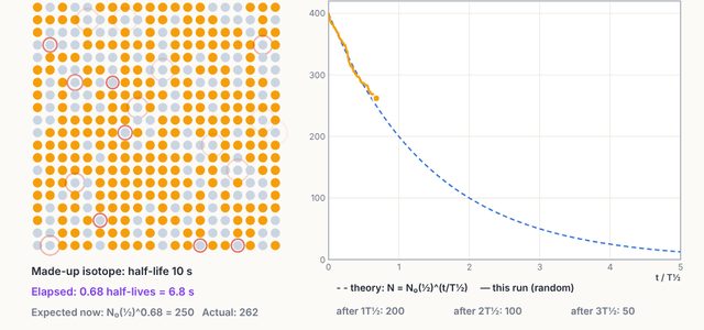
Open the Nuclear Decay & Binding Energy simulation and test every idea on this page yourself.
Open the simulation →